
The 3 biggest pitfalls with using MRI for spinal ligament injury diagnosis are the following:
1. MRI usually leaves 90% of the possible ligament damage to a spine undiagnosed.
2. MRI does not image the most important derangement pattern found in a spinal ligament injury.
3. MRI does not routinely diagnose the severe upper cervical ligament injury.
Pitfall #1: MRI usually leaves 90% of the possible ligament damage to a spine undiagnosed.
To fully understand this first pitfall, look at the anatomy of spinal ligaments and compare to a typical MRI report.
There are 10 ligaments that hold each Spinal Motion Unit together. (There are only 8 ligaments named in the illustration because the capsular ligaments and the transverse ligaments come in pairs, with one on each side.) One of these ligaments is the intervertebral disc, which means the disc is 1/10 or 10% of a Spinal Motion Unit’s ligaments.
No one is ever going to say that the disc is not important, however what about the other 9 ligaments?
To illustrate this, look at a typical MRI report.
The Report starts at C2-3 because that is the first disc space in the human spine
Nowhere is this report are any of the other ligaments mentioned.
MRI has the possibility of being so much more. The machine produces fabulous images but the MRI report content is greatly minimized down to merely a “disc study”. The reports do not address the other 90% of spinal ligament injuries that can exist.
Pitfall #2: MRI does not image the most important derangement pattern with a spinal ligament injury: Excessive Spinal Motion.
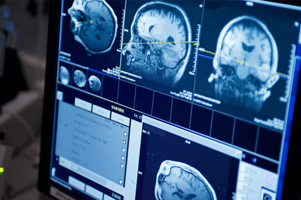
Normal Alignment: when forces that are tolerable act upon spinal ligaments, they remain undamaged; the spine stays in a normal alignment pattern and there is not excessive motion detected. The yellow line on the image is the anterior longitudinal ligament, just 1 of the 10 ligaments that holds a normal Spinal Motion Unit in alignment.
Spinal ligament injury = Misalignment: When spinal ligaments are overcome and damaged by the forces they hyper- stretch, tear or snap in two (avulse) and this always results in excessive motion and instability in the affected joint(s). If the unstable joint is impinging on the spinal cord it causes myelopathy. Impingement of an unstable joint on the spinal nerve causes neuropathy, or it simply causes pain. These are all clinical manifestations of spinal instability. This is excessive spinal motion causing a neurological problem.
Ligaments are tough, flexible tissue that holds bones together to form a joint. It is the job of ligaments to keep all bones in proper position and to allow normal motion in the joints themselves, resulting in proper alignment and proper motion of the joint. When the ligaments are damaged, the bones would start to have excessive joint play or laxity in their movement patterns. This is very easily measured and quantified with special x-rays and an accurate measuring system.
It is not the purpose of an MRI to detect or report on excessive motion in the joints. That is the job of the most inexpensive and accessible imaging that there is which is x-ray, and more importantly stress x-rays.
Pitfall #3: MRI does not routinely diagnose the severe upper cervical ligament injury
Diagnosis means to identify the nature of an illness or injury by assessment of the symptoms (subjective – what the patient complaining of) in conjunction with examination of observable signs (objective – what can be measured, seen, etc.). Nothing may be more productive of symptoms than those associated with damage to what is called the craniocervical junction. The most common symptoms are neck pain, headaches, migraine headaches, cognitive impairment, and visual disturbances.
The craniocervical junction comprises base of the skull (cranium-head), plus cervical 1 (atlas) plus cervical 2 (axis).
The skull is attached to the body by ligaments. The specialized ligaments of the craniocervical junction must allow for stability, yet allow functional movement of the head upon the body. What makes this area so important is that it is traversed by major blood vessels, lymphatic channels, and a great deal of nervous system tissue (spinal nerves, cranial nerves, and the spinal cord). The spacing is tight. Everything is held in alignment (proper spacing) because of these ligaments.
There are two major joints systems in this junction: the atlantooccipital (head to C1atlas) and the atlantoaxial (C1 atlas to C2 axis) joints. These joint systems are extremely different. The atlantoaxial joints allow for about half of the necks range of motion in flexion and extension, but almost no rotational movement. The atlantoaxial joint allows for little flexion and extension movement, but accounts for 25-50% of right and left rotation.
This area is being intensively researched at this time because there is so much neurology, blood flow, lymphatic flow, and cerebral spinal fluid flow associated with this small piece of bodily real estate. Everything that is important for the communication of the head to the body goes through this small area. It is all kept attached, separated, and in alignment by a series of small tough ligaments, called the ligaments of the craniocervical junction. The ligaments we are talking about are everything that is above the C2-3 disc space. Since there is no disc found in this area it is almost never mentioned in a standard cervical MRI report.
The procedure of measuring things in x-rays is very old, probably as old as x-rays themselves. Today measuring excessive joint motion on x-rays of the spine is perhaps one of the most important procedures that can be done. Yet there are very few radiologists that perform this, for one very important reason: they do not have a computer program that allows them to do it accurately.
Spinal Kinetics, LLC that has developed its own technology for doing just that. The process of accurately measuring spinal motion, is called Computerized Radiographic Mensuration Analysis or CRMATM for short. This is quite a simple process in a very complicated field of imaging. Images today come in different formats (arrangement of form), at different outputs and are shot at different distances. These factors and many more are important for capturing good quality films, and can affect the ability to get an accurate read. Therefore, Spinal Kinetics developed their own system for their Board-Certified medical radiologists to use the CRMATM program on any set of spinal x-rays taken in any state in the United States of America.
Accurately detecting excessive spinal motion is even more powerful in the setting of positive findings of a disc herniation on an MRI. When a patient has a disc herniation the first thing their provider should be thinking about is “how bad is the excessive motion”?” A disc herniation along with damage of any of the 9 other ligaments (comprising a spinal motion unit) can result in excessive motion at the same spinal motion unit or at the units above or below the disc herniation. This is a serious, complicated soft tissue injury.
To summarize the 3 pitfalls with MRI and spinal ligament injury diagnosis:
- There are 10 ligaments that hold a spinal motion unit together (disc is one of them) and yet the MRI report usually only mentions damage to the disc and nothing else. MRI is generally only reporting on 1/10 of the spinal ligament structure, which leaves the other 90% undiagnosed.
- MRI is not designed to tell us how much excessive motion damage (instability) the spine has taken from the injury. This omits perhaps the most important finding, as it begins to tell us a lot about the treatments that would be most beneficial and it gives us a great understanding of the time involved with treatment, the expected outcomes and it gives us an early indication of the follow up supportive care the patient may require to maintain the gains from treatment. A disc herniation can be a serious injury, however a disc herniation with no excessive joint movement and one with severe joint movement are two completely different disc herniation’s!
- MRI does not routinely diagnose the severe upper cervical ligament injury
Conclusion
Spinal ligament injuries are some of the most significant injuries that any human body can receive, simply because they put the patient at such high risk for long-term residual complaints. Often these complaints are not accompanied by accurate documentation of physical findings, simply because the doctors that are working with these patients are not aware of what such injuries look like.
In this report, I chose not to focus on such things as MRI signal intensities to pick up ligament damage or other such complicated and often variable procedures. These injuries can be very easily picked up with standard stress radiology there was no need to get any more complicated.
All problems are resolved by applying basics. The purpose of this report was to bring to the forefront the simple imaging procedures that are not being standardly applied today.
Every patient suspected of having a spinal ligament injury should be tested with simple stress radiology and a CRMATM excessive spinal motion study. I hope that this report was straightforward enough to provide you with a great understanding of why. When patients are not in the hands of doctors that understand their injuries, their clinical outcomes obviously can be questionable. Time matters in these types of injuries, as every day the patient goes without a clear understanding of what they have the greater the risk the patient can wind up with a symptom that becomes permanent. Spinal ligament injuries are permanent, but the symptoms produced do not have to be.
References:
1. http://www.lanjochiro.com/2012/08/what-is-forward-head-posture/
2.https://crashstats.nhtsa.dot.gov/Api/Public/ViewPublication/812013
3. https://www.pbacloudb2019.com/
4. https://americanpainsociety.org
5. https://www.sciencedaily.com/releases/2012/09/120911091100.htm